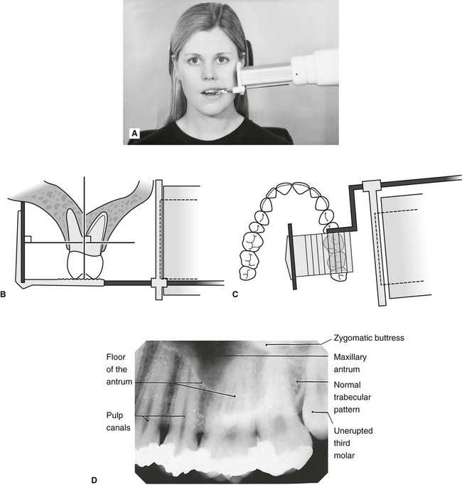
Free Shipping On Orders Over 70. With this technique the film is placed parallel to the long axis of a tooth allowing the X-ray to be focused perpendicular to the long axis of the tooth.
Periapical Radiography Pocket Dentistry
Ensure they are seated high enough so it is easy to see the occlusal.

. Because the film is placed in the mouth at an angle to the long axis of the teeth. Periapical X-rays show the entire tooth from the exposed crown to the end of the root and the bones that support the tooth. The snap-a-ray is used.
Instruction is provided in the use of dent hammers dent balls and barrels mandrels burnishers and other tools of the industry. The paralleling technique results in good quality x-rays with a minimum of distortion and is the most reliable technique for taking periapical x-rays. I Periapical X-ray corroborates the periodontal regeneration in close contact with MTA filling.
Periapical x-ray is a type of x-ray that shows one to two teeth from their crown to their root and the surrounding bone. Parallel technique The image receptor is placed in a holder and placed in the mouth parallel to the longitudinal axis of the tooth under. Size 2 Film is used for Anterior and Posterior X-rays when Bisecting.
Since the slope and curvature of the dental arches and the alveolar processes will not permit the film to be held close to the teeth. When comparing the two periapical techniques the. Periapical views are used to record the crowns roots and surrounding bone.
X ray films hmdali. The patient is seated upright in the dental chair and should remove any removable dental appliances glasses or jewelry that could interfere with the X-ray beam. Single periapical radiographs are often made of individual teeth or groups of teeth to obtain information for treatment or diagnosis of localized diseases or abnormalities.
Periapical film is held parallel to the long axis of the tooth using film-holding instruments. The image receptor is placed in a holder and positioned in the mouth parallel to the long axis of the tooth under. Since the slope and curvature of the dental arches and the alveolar processes will not permit the film to be held close to the teeth.
To take a periapical exposure the hygienist or x-ray technician places a small photosensitive imaging plate coated with phosphorus into a sterile wrapper and inserts it into the patients mouth just like a conventional X-ray film card. Paralleling technique Bisecting angle technique Paralleling technique It is also called the extension cone paralleling technique right angle technique and long cone. The X-ray is taken and the exposed plate is then loaded into a scanner or processor which reads the image.
Ad Buy Dental Supplies Online At Safco Dental Supply. Paralleling Technique for Periapical X-rays The paralleling technique results in good quality x-rays with a minimum of distortion and is the most reliable technique for taking periapical x-rays. The film is placed parallel to the long axis of the tooth in question and the central x-ray beam should be directed perpendicular to the long axis of the tooth.
The bisecting short-cone and paralleling long-cone techniques are two of the most commonly used techniques. Paralleling Technique for Periapical X-rays The paralleling technique results in good quality x-rays with a minimum of distortion and is the most reliable technique for taking periapical x-rays. Periapical radiographs provide important information about the teeth and surrounding bone.
Most frequently used radiography is for the periapical which is performed by the bisecting Thus when considering the execution of the radiographic technique and the possibility of errors that occur during the exposure of X-ray image XR receptors it is important to identify those that occur more frequently. Periapical X-rays are used to detect any abnormalities of the root structure and surrounding bone structure. Extraoral radiograph Panoramic X-ray Tomograms Cephalometric projections Sialography Computed tomography 10.
These X-rays are used to find dental problems below the gum line or in the jaw such as impacted teeth tooth fractures abscesses tumours and bone changes linked to some diseases. The film is placed parallel to the long axis of the tooth to be radiographed and the central beam of X-ray is directed at right angle to the film and the teeth. Occlusal X-rays show full tooth development and placement 9.
Different techniques and instruments are used to drain and decompress large periapical lesions ranging from placing a stainless steel tube into the root canal exhibiting persistent apical exudation 202 204 which is non-surgical decompression to placing polyvinyl or polyethylene tubes through the alveolar mucosa covering the apical lesion which is surgical. The snap-a-ray is used. The film is placed parallel to the long axis of the tooth in question and the central x-ray beam should be directed perpendicular to the long axis of the tooth.
The X-ray tube head should be positioned so that the beam meets the tooth and the image. Low Prices Quality Service Every Day. The extraoral periapical radiographic technique was performed for both maxillary and mandibular teeth using Newman and Friedman technique2.
A long cone is used to take x-rays with paralleling exposure techniques. The central ray is directed to pass at a perpendicular angle to both the tooth and the film. By using a filmsensor holder with fixed image receptor and.
By using a film sensor holder with still. Periapical radiography is a commonly used intraoral imaging technique in radiology and may be a component of your radiologic examination. For this purpose a special technique of periapical radiography was developed by Gordon M.
The X-ray head is directed at right angles vertically and horizontally of both the tooth and the image receptor. Periapical film is held parallel to the long axis of the tooth using film-holding instruments. When comparing the two periapical techniques the advantages of the bisecting angle technique are.
The patient was positioned upright with hisher mouth was opened as wide as possible to allow the X-ray beam to pass to the sensor unobstructed from the opposite side of the mouth. Fitzgerald called as paralleling or long cone technique. The X-ray tubehead is then aimed at right angles vertically and horizontally to both the tooth and the image.
This type of x-ray can be used for any tooth in the mouth and it is mostly used to determine the depth of the decay and if the tooth needs endodontic therapy if there are any. The paralleling technique results in good quality x-rays with a minimum of distortion and is the most reliable technique for taking periapical x-rays. Basically there are two techniques for taking periapical radiography.
Periapical X-rays. 30 Day Return Policy. Both techniques have advantages and disadvantages.
It is a very commonly used diagnostic measure.

Periapical Radiography Pocket Dentistry

Periapical Radiography Pocket Dentistry

Periapical Radiography Pocket Dentistry
Periapical Radiography Pocket Dentistry

Periapical Radiography Pocket Dentistry

Periapical Radiography Pocket Dentistry

Periapical Radiography Pocket Dentistry

How Make Periapical X Ray
0 comments
Post a Comment